|
Muscle
|
 Click on image for larger view |
|
|
Name
|
Quadriceps Femoris
|
|
|
Subdivision
|
vastus lateralis
|
|
|
Muscle Anatomy
|
||
|
Origin
|
Proximal parts of intertrochanteric line, anterior and inferior borders of greater trochanter, lateral lip of gluteal tuberosity, proximal half of lateral lip of linea aspera, and lateral intermuscular septum. | |
|
Insertion
|
Proximal border of the patella and through patellar
ligament.
|
|
|
Function
|
Extension of the knee joint.
|
|
|
Recommended sensor placement procedure
|
||
|
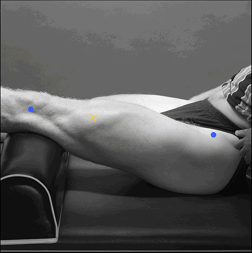
Starting posture
|
Sitting on a table with the knees in slight flexion
and the upper body slightly bend backward.
|
|
|
Electrode size
|
Maximum size in the direction of the muscle fibres:
10 mm.
|
|
|
Electrode distance
|
20 mm.
|
|
|
Electrode placement
|
||
|
- location
|
Electrodes need to be placed at 2/3 on the line
from the anterior spina iliaca superior to the lateral side of the
patella.
|
|
|
- orientation
|
In the direction of the muscle fibres
|
|
|
- fixation on the skin
|
(Double sided) tape / rings or elastic band.
|
|
|
- reference electrode
|
On / around the ankle or the proc. spin. of C7.
|
|
| Clinical test | Extend the knee without rotating the thigh while applying pressure against the leg above the ankle in the direction of flexion. | |
| Remarks |
The SENIAM guidelines include also a separate sensor
placement procedure for the vastus medialis and the rectus femoris
muscle.
|
|