|
Muscle
|
||
|
Name
|
Gluteus
|
|
|
Subdivision
|
Maximus
|
|
|
Muscle Anatomy
|
||
|
Origin
|
Posterior gluteal line of ilium ad portion of bone superior and posterior to t, posterior surface of lower part of sacrum, side of coccyx, aponeurosis of erector spinea, sacrotuberous ligament and gluteal aponeurosis. | |
|
Insertion
|
Larger proximal portion and superficial fibres of
distal portion of muscle into iliotibial tract of fascia lata. Deeper
fibres of distal portion into gluteal tuberosity of femur.
|
|
|
Function
|
Extends, laterally rotates and lower fibres assist
in adduction of the hip joint. The upper fibres assist in adduction.
Through its insertion into the iliotibial tract, helps to stabilise
the knee in extension.
|
|
|
Recommended sensor placement procedure
|
||
|
Starting posture
|
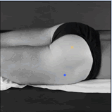
Prone position, lying down on a table.
|
|
|
Electrode size
|
Maximum size in the direction of the muscle fibres:
10 mm.
|
|
|
Electrode distance
|
20 mm.
|
|
|
Electrode placement
|
||
|
- location
|
The electrodes need to be placed at 50% on the line
between the sacral vertebrae and the greater trochanter. This position
corresponds with the greatest prominence of the middle of the buttocks
well above the visible bulge of the greater trochanter.
|
|
|
- orientation
|
In the direction of the line from the posterior
superior iliac spine to the middle of the posterior aspect of the
thigh
|
|
|
- fixation on the skin
|
(Double sided) tape / rings or elastic band.
|
|
|
- reference electrode
|
On the proc. spin. of C7 or on / around the wrist
or on / around the ankle.
|
|
| Clinical test | Lifting the complete leg against manual resistance. | |
| Remarks |
The SENIAM guidelines include also a separate sensor
placement procedure for the gluteus medius muscle.
|
|
