|
Muscle
|
||
|
Name
|
Biceps femoris
|
|
|
Subdivision
|
Long head and short head
|
|
|
Muscle Anatomy
|
||
|
Origin
|
Long head: distal part of sacrotuberous ligament and posterior part
of tuberosity Short head: lateral lip of linea aspera, proximal 2/3 of supracondylar line and lateral intermuscular septum. |
|
|
Insertion
|
Lateral side of head of fibula, lateral condyle
of tibia, deep fascial on lateral side of leg.
|
|
|
Function
|
Flexion and lateral rotation of the knee joint.
The long head also extends and assists in lateral rotation of the
hip joint.
|
|
|
Recommended sensor placement procedure
|
||
|
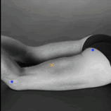
Starting posture
|
Lying on the belly with the face down with the thigh
down on the table and the knees flexed (to less than 90 degrees)
with the thigh in slight lateral rotation and the leg in slight
lateral rotation with respect to the thigh.
|
|
|
Electrode size
|
Maximum size in the direction of the muscle fibres:
10 mm.
|
|
|
Electrode distance
|
20 mm.
|
|
|
Electrode placement
|
||
|
- location
|
The electrodes need to be placed at 50% on the line
between the ischial tuberosity and the lateral epicondyle of the
tibia.
|
|
|
- orientation
|
In the direction of the line between the ischial
tuberosity and the lateral epicondyle of the tibia.
|
|
|
- fixation on the skin
|
(Double sided) tape / rings or elastic band.
|
|
|
- reference electrode
|
On / around the ankle or the proc. spin. of C7.
|
|
| Clinical test | Press against the leg proximal to the ankle in the direction of knee extension. | |
| Remarks | ||
